It's easy to feel powerless against cancer, but a new study has identified several ways that we can reduce the odds of it occurring.
According to new analysis from the World Health Organization (WHO), more than a third of all cancer cases globally are preventable.
Lung, stomach, and cervical cancers make up nearly half of those cases.
This means that millions of deadly cancers every year could be prevented through medical intervention, behavior changes, reducing occupational risks, or tackling environmental pollutants.
"Addressing these preventable causes represents one of the most powerful opportunities to reduce the global cancer burden," says Isabelle Soerjomataram, medical epidemiologist at WHO and senior author of the analysis.
The analysis found that in 2022, there were nearly 19 million new cases of cancer. Roughly 38 percent of those diagnoses were related to 30 changeable risk factors.
These included tobacco smoking, alcohol consumption, high body mass index, insufficient physical activity, smokeless tobacco (like chewing tobacco), a traditional stimulant known as areca nut, suboptimal breastfeeding, air pollution, ultraviolet radiation, infectious agents, and over a dozen occupational exposures.
The number one preventable factor associated with cancer? Smoking tobacco. It was linked to 15 percent of all cancer cases that year.
For men, the risk was particularly high. Smoking contributed to 23 percent of all new cancer cases globally in men that year.
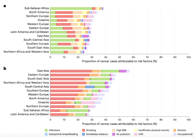
But smoking isn't the only cause; air pollution also plays a role, and its impact varies by region. In East Asia, for instance, about 15 percent of all lung cancer cases in women were due to air pollution. In Northern Africa and Western Asia, meanwhile, approximately 20 percent of all lung cancer cases in men were due to air pollution.
After tobacco smoking, the runner-up among changeable lifestyle factors was drinking alcohol. It accounted for 3.2 percent of all new cancer cases (approximately 700,000 cases).

Infections, meanwhile, were linked to roughly 10 percent of new cancer cases. Among women, the largest share of preventable cancers was due to high-risk human papillomavirus (HPV), which can lead to cervical cancer.
Thankfully, we now have a vaccine for HPV that prevents many of these associated diseases, and yet coverage in many parts of the world remains low.
Stomach cancer cases are higher among men and tend to be associated with smoking and infections due to overcrowding, inadequate sanitation, and poor access to clean water.
Related: US Cancer Survival Has Reached a Milestone High of 70%
"By examining patterns across countries and population groups, we can provide governments and individuals with more specific information to help prevent many cancer cases before they start," says André Ilbawi, WHO Team Lead for Cancer Control and co-author of the analysis.
Now it's time to roll up our sleeves.
The study was published in Nature Medicine.
]]>
A common bacterium usually found in the respiratory system appears to be linked to cognitive decline and Alzheimer's disease when it's present in the retina.
Chlamydia pneumoniae – often responsible for pneumonia and sinus infections – has previously been spotted in brains affected by Alzheimer's. Now, a new study has detected C. pneumoniae in the vision-generating tissue that lines the back of the eye, at higher levels in people with Alzheimer's.
Led by a team from Cedars-Sinai Medical Center in the US, the research provides fresh insight into the biological processes that may worsen Alzheimer's progression – and could inspire new approaches to slowing the disease.
As well as potentially contributing to the cascade of mechanisms that lead to Alzheimer's, the presence of C. pneumoniae in the retina could also one day be used to detect cognitive decline and dementia – though that possibility wasn't directly tested here.
"The eye is a surrogate for the brain, and this study shows that retinal bacterial infection and chronic inflammation can reflect brain pathology and predict disease status, supporting retinal imaging as a noninvasive way to identify people at risk for Alzheimer's," says neuroscientist Maya Koronyo-Hamaoui, from the Cedars-Sinai Medical Center.
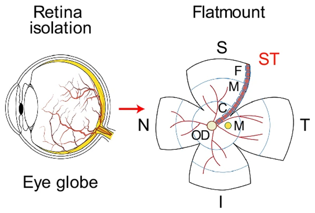
To begin with, the team analyzed eye and brain tissue from 104 people after death. Some had Alzheimer's disease, some had mild cognitive impairment (MCI), and some hadn't reported any cognitive problems.
They found a clear association between the presence of C. pneumoniae in the eye and brain and having a diagnosis of Alzheimer's. Higher levels of the bacterium in tissue were linked to more severe cognitive decline.
People with APOE gene variants linked to Alzheimer's risk also had higher levels of the bacterium in their tissues. However, the differences between people without cognitive impairment and those with MCI were much less clear-cut when it came to C. pneumoniae.
Next, the researchers ran tests using lab-grown neurons and animal models to determine what C. pneumoniae might be doing biologically. These experiments showed that infections with the bacterium led to increased inflammation, greater cognitive decline, and more nerve cell death.
The presence of C. pneumoniae was also associated with increased amounts of amyloid-beta protein in the brain, which is known to clump together in dangerous ways in the brains of people with Alzheimer's.
"Seeing Chlamydia pneumoniae consistently across human tissues, cell cultures, and animal models allowed us to identify a previously unrecognized link between bacterial infection, inflammation, and neurodegeneration," says Koronyo-Hamaoui.
There are still unanswered questions, and the findings are only a strong suggestion that C. pneumoniae could contribute to (and be a sign of) Alzheimer's disease – not conclusive proof.
However, if infection by the bacterium is indeed leading to inflammation that extends to the brain and accelerates neurodegenerative processes, then we may have a new target for future treatments.
The researchers describe C. pneumoniae as a potential amplifier rather than a primary trigger, which aligns with growing evidence of just how complex Alzheimer's is. It's likely there are multiple contributing factors that may differ between people.
What's more, the team identified a specific inflammation pathway that C. pneumoniae targets, possibly worsening the damage already being done by Alzheimer's. Further studies will be required to confirm this mechanism, but the signs are there.
Related: A Common Sleeping Pill May Reduce Buildup of Alzheimer's Proteins, Study Reveals
Scientists continue to identify multiple ways the eyes and the brain are linked. In this case, the findings could prove valuable to society's ongoing efforts to combat Alzheimer's and other forms of dementia.
"This discovery raises the possibility of targeting the infection-inflammation axis to treat Alzheimer's," says biomedical scientist Timothy Crother, from the Cedars-Sinai Medical Center.
The research has been published in Nature Communications.
]]>
Physical abilities fade as we age, but many of us like to think that won't be an issue until we're well into our golden years.
According to a new study, however, fitness and strength begin to dwindle as early as age 35, regardless of exercise habits. This is followed by a gradual decline that accelerates with age.
While this fate may be unavoidable, that doesn't mean it's out of our hands. Even if physical activity won't help us delay our peak, it can make a difference in how rapidly our abilities deteriorate, the study suggests.
Aging involves a progressive decline in skeletal muscle, which can noticeably manifest for some people in their 60s, sometimes limiting mobility.
Previous research on elite athletes has shown that, despite continuous training, physical performance typically peaks by about age 30. This suggests the mechanics behind age-related muscle loss could already be at work decades before they become clinically significant.

There are advantages to studying physical abilities in athletes, such as data availability and lack of interference by sedentary lifestyles, but there is also the "obvious disadvantage" that elite athletes may not be representative of the general population, the authors note.
For the new paper, researchers conducted a population-based longitudinal study in hopes of measuring the physical capacity of the general population from adolescence to older age.
Research on this subject has typically relied on cross-sectional studies, which analyze data from a population at a specific point in time. Longitudinal studies can therefore provide valuable perspectives on how variables may change over a period.
The researchers used data from the Swedish Physical Activity and Fitness (SPAF) cohort study, a longitudinal study that has been following several hundred participants in Sweden since 1974, when they were 16 years old.
The SPAF includes strength and fitness data from these same people at five intervals in the last five decades (ages 16, 27, 34, 52, and 63), offering a unique opportunity to measure changes in physical abilities over half a century.
Cross-sectional studies seem to have underestimated the age-related decline in physical capacity, the researchers report, but their findings support existing evidence that it affects men and women similarly.
For both sexes, muscular endurance and estimated maximal aerobic capacity peaked between ages 26 and 36 before gradually declining, first by 0.3 percent to 0.6 percent per year, and later by up to 2.5 percent per year, with no sex difference in the rate of decline.
There was a difference in muscle power, with men and women peaking at ages 27 and 19, respectively. Their muscle power then faded at similar rates, initially decreasing by 0.2 percent to 0.5 percent per year, and later escalating to an annual decline of 2 percent or more. By age 63, participants' overall drop from their peak physical capacity ranged from 30 percent to 48 percent.
There is good news. While we may be unable to dodge or delay our physical decline, we can reduce its speed with regular exercise, the authors report.
"Individuals who were physically active in their leisure time at age 16 maintained higher aerobic capacity, muscular endurance, and muscle power throughout the observation period," they write.
This highlights the importance of promoting physical activity to teenagers and young adults, but that message is helpful no matter how old you are. Participants who became more active in adulthood still managed to improve their physical capacity by around 10 percent, the study found.
Related: Study Reveals The Surprising Age at Which Your Brain Reaches Its Peak
"It is never too late to start moving. Our study shows that physical activity can slow the decline in performance, even if it cannot completely stop it," says lead author Maria Westerståhl, lecturer in the Department of Laboratory Medicine at the Karolinska Institute.
"Now we will look for the mechanisms behind why everyone reaches their peak performance at age 35, and why physical activity can slow performance loss but not completely halt it," Westerståhl says.
The study was published in the Journal of Cachexia, Sarcopenia and Muscle.
]]>
Yawning has an unusual and unexpected effect on the flow of fluid protecting the brain, a recent study reveals, though it's not yet clear what the impact of this shift might be.
According to researchers from the University of New South Wales in Australia, the findings could provide a crucial clue in understanding why humans (and many other species) evolved the capacity to yawn.
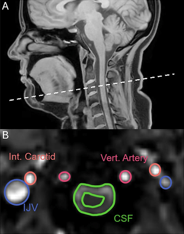
The research team used MRI to scan the heads and necks of 22 healthy participants while they were told to yawn, take deep breaths, stifle yawns, and breathe normally.
Given that yawning and deep breathing share similar mechanisms, the researchers expected them to look similar on the scans. Surprisingly, the images revealed a key difference: unlike deep breaths, yawns sent cerebrospinal fluid (CSF) away from the brain.
"The yawn was triggering a movement of the CSF in the opposite direction than during a deep breath," neuroscientist Adam Martinac told James Woodford at New Scientist.
"And we're just sitting there like, whoa, we definitely didn't expect that."
This wasn't observed in every case, and occurred less often in men, though the researchers caution that this may be due to interference from the scanner itself.
The analysis also revealed that both deep breaths and yawns increased the flow of blood leaving the brain, making more room for fresh blood to be pumped in.
Blood flow didn't change direction with yawns. Yet during its initial stages, carotid arterial blood flow into the brain surges by around a third, providing potential evidence for multiple reasons for the behavior.
In addition, the participants all had unique yawning patterns that were closely followed each time they yawned. It's a sign that we all have our own central pattern generator determining how we yawn.
"This flexibility might account for the variations in inter-participant yawning patterns while still maintaining a recognizable, individual-specific pattern; and implies that the patterns of yawning are not learned but are an innate aspect of neurological programming," write the researchers in their paper.
The next big question is what all of this means, and why yawns should differ from deep breaths so substantially when it comes to CSF, a fluid that keeps the central nervous system running smoothly, delivering nutrients and removing waste.
One possibility raised by the researchers is that yawning has a specific role in cleaning out the brain. Another idea is that it's some kind of brain cooling function in operation.
Yawns do appear to be closely connected to the brain and the central nervous system – bigger brains typically lead to longer yawns, for example, perhaps a nugget of trivia you can share with friends and family the next time you yawn for an extended period of time.
Related: This Article on The Science of Yawning Will Probably Make You Yawn
Yawning continues to be a rather baffling phenomenon with a largely unclear purpose, despite being a behavior seen in many different species, and which tends to be contagious among people and animals.
"Yawning appears to be a highly adaptive behavior and further research into its physiological significance may prove fruitful for understanding central nervous system homeostasis," write the researchers.
The research has yet to be peer-reviewed, but is available on bioRxiv.
]]>
For more than a century, the Green River's course through the Uinta Mountains in Utah's northeast has been a geological mystery, seemingly defying physics.
Rivers carve their paths by flowing downhill across many years, which means they usually follow the slopes and furrows of any mountain ranges they encounter.
And yet the Green River, which has been following this course for just 8 million years, cuts right across the 50-million-year-old mountains to meet with the Colorado River, etching out the 700-meter (roughly 2,300-foot) deep Canyon of Lodor that runs perpendicular to the range (and all logic).
Geologist Adam Smith from the University of Glasgow in Scotland led a team to interrogate this long-held mystery. It turns out, the Green River did not have to flow uphill at all: instead, the mountain range was conveniently lowered, in a phenomenon known as lithospheric drip.

"Other rivers in the Uinta Mountains provide evidence that the height of the Uinta Mountains changed in the last few million years," Smith and team write.
Their data suggests the root of the Uinta Mountains, a dense mineral chunk at the base of the lithosphere, became so heavy that it 'dripped' into Earth's liquid mantle. This would have temporarily pulled the mountain range down, allowing the Green River to chart its unlikely course.
Later, the Uinta mountains grew by 400 meters up around the river, forming the canyon we have today.
Seismic imaging involves reading the scatter of earthquake vibrations as they pass through Earth to create a picture of what's going on down there. At the Uinta Mountains, seismic images revealed a cold, round chunk about 200 kilometers below the surface: probably the drip in question.
What's more, the crust below these mountains is much thinner than you'd usually expect: more evidence that the drip had torn away the lower layers.
Related: Earth's Crust Is Dripping Under Midwest US, Scientists Discover
Once this drip broke free from the lithosphere about 2-5 million years ago, the mountain range was able to rebound. By then, the Green River had settled in for good: The Canyon of Lodor was there to stay, and the Green River became a tributary of the Colorado River.
"The merging of the Green and Colorado Rivers millions of years ago altered the continental divide of North America," Smith explains.
"It created the line that separates the rivers that flow into the Pacific from those that flow into the Atlantic, and created new habitat boundaries for wildlife that influenced their evolution."
The research is published in the Journal of Geophysical Research: Earth Surface.
]]>
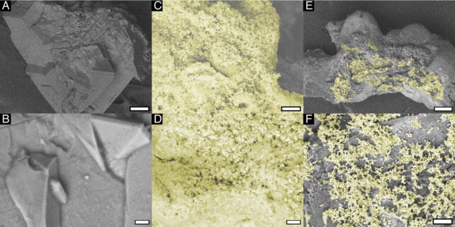
For the first time, scientists have discovered evidence of bacteria hiding in 'noninfectious' kidney stones.
These hardened clumps of small crystals are made from chemicals in urine and are thought to form due to a lack of fluid or a high concentration of minerals and chemicals.
Most kidney stones are considered noninfectious after they are passed. But that may not be true after all.
A study led by researchers at the University of California Los Angeles (UCLA) has now found that calcium oxalate kidney stones, the most common type, are enriched with bacteria.
In fact, these stones contain sheets of bacteria as part of their intrinsic internal structure.
"This breakthrough challenges the long-held assumption that these stones develop solely through chemical and physical processes, and instead shows that bacteria can reside inside stones and may actively contribute to their formation," explains urologist Kymora Scotland from UCLA.
"By uncovering this novel mechanism, the study opens the door to new therapeutic strategies that target the microbial environment of kidney stones."
The findings suggest that in some cases where kidney stones keep coming back, there may be a bacterial infection in the kidney, ureter, or bladder contributing to recurrence.
If this infection is treated, then perhaps there may be fewer kidney stones going forward.
Using electron and fluorescence microscopy, the researchers found structural and chemical evidence of bacteria in calcium oxalate stones taken from human patients.
This was true even among participants without underlying urinary tract infections.
"We found a new mechanism of stone formation that may help to explain why these stones are so common," says Scotland.
"These results may also help to explain the connections between recurrent urinary tract infections and recurrent kidney stone formation, and provide insights on potential future treatment for these conditions."
Related: These 7 Common Daily Habits Could Be Damaging Your Kidneys
As many as one in 11 people will suffer from kidney stones in a lifetime, and more than 70 percent of cases are attributed to calcium oxalate stones.
If bacteria play a significant role in their formation, then treatments and guidance for kidney stones may need to be updated.
The study was published in PNAS.
]]>
Could humanity nuke an incoming asteroid to deflect it and save the Earth, disaster-movie style? A unique new impact simulation suggests that a nuclear option could be a viable last resort to avert an apocalypse.
Researchers have recently found that space rocks can withstand much more stress than previously inferred from experiments and observations. Counter-intuitively, asteroids actually grow stronger when subjected to an intense impact.
It may sound discouraging, but this discovery can improve planetary defense strategies because it suggests that a nuked asteroid will remain intact, rather than fragmenting into many space rocks that would rain down across our planet.
As detailed in a recently released paper, a team of researchers, including physicists from the University of Oxford, partnered with the Outer Solar System Company (OuSoCo), a nuclear deflection startup, to analyze what happens to an iron space rock under different levels of stress.
"These analyses are intended to examine changes in the meteorite's internal structure caused by the irradiation and to confirm, at a microscopic level, the increase in material strength by a factor of 2.5 indicated by the experimental results," explains Melanie Bochmann, co-founder of OuSoCo and co-leader of the research team.

Like the DART mission displayed in 2022, one promising way to avert an asteroid-induced apocalypse is to deflect the incoming threat with a kinetic impactor, a human-made cosmic battering ram sent to smash into a looming asteroid at many times the speed of a bullet.
It's conceptually simple, but the reality is fraught with perilous uncertainties; a hit in the wrong spot may only delay an asteroid's doomsday approach toward Earth. Furthermore, the impactor's energy and the asteroid's material response can lead to unexpected consequences like fragmentation or a surprising shift in momentum.
So, to decide between an impactor like DART or an as-yet untried nuclear approach, planetary defenders must ascertain the mechanical behavior of different asteroid materials. This knowledge is essential to transfer energy to said asteroid and redirect its trajectory away from Earth.
Yet such data is scarce, especially data that shows how materials react in real-time. For example, different models yield different values for yield strength, a measure of how easily a body breaks under stress.
These models may differ by up to a factor of seven, depending on whether they test for local (microscopic) or bulk (macroscopic). Additionally, the destructive nature of previous tests precluded direct measurement of material responses as they occurred.
"This is the first time we have been able to observe – non-destructively and in real time – how an actual meteorite sample deforms, strengthens, and adapts under extreme conditions," says Gianluca Gregori, a physicist at the University of Oxford and one of the study's co-authors.
Researchers employed a unique technique to ensure they didn't destroy the evidence. They used the Super Proton Synchrotron particle accelerator at CERN's High Radiation to Materials (HiRadMat) facility to irradiate a sample from a Campo del Cielo iron meteorite, blasting it with high-energy, short-duration proton beam pulses at lower and higher intensities.
As a result, temperature sensors and laser Doppler vibrometry (a technique to analyze surface vibrations) revealed that the meteorite sample softened, flexed, then surprisingly re-strengthened. It also displayed a quality called strain-rate dependent damping, which means that the harder it's hit, the more effectively it dissipates energy.
This study method provides invaluable data that explain why discrepancies in yield strength observed in previous laboratory experiments differ from evidence of meteor fragmentation in Earth's atmosphere, and that these discrepancies are due to factors such as internal stress redistribution.
It also highlights that these mechanical properties evolve in real time and should not be assumed to be fixed, as may often be the case in existing asteroid deflection models. Further research will involve other types of asteroid compositions.
Here, researchers chose an iron-rich sample for its relative homogeneity, but more heterogeneous space rocks will exhibit different stress-dissipation capabilities based on the spatial distribution of their constituent materials.
The ultimate scope of this research will hopefully remain theoretical:
"The world must be able to execute a nuclear deflection mission with high confidence, yet cannot conduct a real-world test in advance. This places extraordinary demands on material and physics data," says Karl-Georg Schlesinger, co-founder of OuSoCo and co-leader of the research team.
Related: NASA: Nuclear Explosion Could Save Moon From Asteroid Strike in 2032
However, should a nuclear option ever be necessary, it likely won't mirror the movies – no drilling necessary. Instead of loading an asteroid with explosives, some physicists propose a standoff nuclear detonation near an asteroid to vaporize part of its bulk and deflect its orbital path.
This research is published in Nature Communications.
]]>
Supplementing the guts of older mice with poop from younger ones has revealed the key role microbes play in intestinal stem cell function.
After receiving a fecal microbiota transplant from younger mice, one aspect of age-related decline in the guts of older mice was reversed, driven by increased intestinal stem cell activity that maintains the intestinal walls.
The findings suggest that such transplants could someday be a treatment pathway for age-related intestinal conditions, such as inflammation and obesity.
"As we age, the constant replacement of intestinal tissue slows down, making us more susceptible to gut-related conditions," says molecular biologist Hartmut Geiger of Ulm University in Germany. "Our findings show that younger microbiota can prompt older intestine to heal faster and function more like younger intestine."
Intestinal stem cells are crucial for maintaining a stable, healthy gut. They're the mechanism by which the gut lining – the epithelium – constantly replenishes and renews itself, ensuring consistent gut function.
However, as we age, the rate of this renewal slows, increasing vulnerability to age-related gut dysfunction.
In previous research, Geiger and his colleagues, cell biologists Yi Zheng and Kodandaramireddy Nalapareddy of Cincinnati Children's Hospital Medical Center, determined that this slowed regeneration is directly linked to reduced function of intestinal stem cells.
We also know that the microbial communities that live in our guts change with age, with such changes linked to conditions like Parkinson's disease, Alzheimer's disease, and even vision loss. The researchers wanted to know if the gut microbiome affects stem cell activity, too.
So, they recruited more team members and designed a simple experiment to test it: transplanting fecal samples between and within groups of old mice and young mice.
After the series of transplants was complete, the researchers studied the intestines to see what changes, if any, resulted from the transfer.
In the older mice, the change was dramatic. Stem cell activity had increased, as well as the Wnt signalling that these cells need in order to function. The regeneration of the epithelium picked up pace – and, critically, the gut healed more quickly after radiation damage.
"This reduced signaling causes a decline in the regenerative potential of aged ISCs," Zheng says. "However, when older microbiota were replaced with younger microbiota, the stem cells resumed producing new intestine tissue as if the cells were younger. This further demonstrates how human health can be affected by the other life forms living inside us."
In the younger mice, the change was less dramatic. There was only a slight drop in stem cell activity, Wnt signalling, and regeneration; the intestines continued to function reasonably well. This suggests that the aging gut is far more sensitive to microbiome changes than younger ones.
Another really interesting finding was that one of the perpetrators of stem cell curtailment in the aging gut is Akkermansia, a bacterium that is generally considered beneficial in several ways, with signs that it can help reduce diet-induced obesity and depression-like behavior in mice.
In aging mice, elevated levels of Akkermansia actually contribute to the suppression of Wnt signalling – a finding that implies that gut bacteria are not necessarily good or bad, but that their contribution may depend on context.
This isn't a slam-dunk for human health; our bodies (and intestines) are more complex than those of mice, and we'd need to perform separate studies to see if this phenomenon occurs in our own species.
Related: Most People Develop Diverticulosis in Their Gut by Age 80… So What Is It?
However, the research does illuminate a promising avenue for future study.
It also suggests that age-related stem cell decline may not be irreversible. By harnessing the ability of gut microbes to shape how intestinal tissue renews itself, scientists could one day develop ways to help preserve intestinal health as we age.
The research has been published in Stem Cell Reports.
]]>
'Zombie' coronavirus fragments not only help drive inflammation in long-COVID, but also destroy our immune cells.
A recent study by an international team of more than 30 authors reveals how the destruction of the virus within our body leaves dangerous protein fragments that target specific immune cells, which may explain some of the debilitating consequences millions of people with long-COVID now face.
"These fragments target a specific kind of curvature on the membranes of cells," explains bioengineer Gerard Wong from the University of California, Los Angeles. "Cells that are spiky, that are star-shaped, or that have lots of tentacles end up getting preferentially suppressed."
These "spiky" cells include early-warning dendritic cells that detect and alert the rest of the body to viruses, as well as CD8+ and CD4+ T cells that help destroy already infected cells.
Previous research already noted this T cell depletion, which has since been recognized as a plausible diagnostic tool.
"Viruses do so many things that we don't understand," says Wong. "We want to understand what all the leftover viral matter does to us, both during COVID and after. With these viral fragments, all of a sudden there's a whole new range of possibilities to consider."
The fact that multiple types of these viral fragments can attack immune cells may explain why those with preexisting immune conditions are more susceptible to these impacts, even when they are otherwise healthy.
In further verification, the Omicron strain of COVID-19, infamous for being highly infectious but less dangerous, breaks down into a greater variety of protein fragments in our bodies than previous strains.
"No one could really explain why it replicated as fast as the original strain but generally did not cause infections that were as serious," explains Yue Zhang, a bioengineer at China's Westlake University.
"We found that pieces of the Omicron spike were much less able to kill these important immune cells – suggesting that a patient's immune system is not going to be as depleted."
Despite the rhetoric of the pandemic being a thing of the past, COVID-19 continues to kill about 100,000 people in the US annually and disable many more. Up to 17 million people in the US had long COVID in 2024.
Related: Strange Structures Found Lurking in The Blood of People With Long COVID
Amid this backdrop, many have been left struggling without adequate support for the ongoing consequences of long COVID, which are very real and debilitating. What's more, recent studies found that the risk of long COVID can increase with subsequent infections in children and adults.
"One of the strongest reasons I give patients, families, and physicians about getting vaccinated: More vaccines should lead to fewer infections, which should lead to less long COVID," urged pediatrician Ravi Jhaveri of Lurie Children's Hospital in Chicago last September.
This research was published by PNAS.
]]>
Putting aside the risk of an early grave by accident or injury, your genes may have a much greater impact on your lifespan than previously thought, according to an unparalleled study of twin data.
The recent analysis, led by researchers from the Weizmann Institute of Science in Israel, suggests that around 55 percent of the variation in human lifespans is influenced by genes.
That's much higher than previous estimates of around 20-25 percent, with measures falling as low as just 6 percent in some studies.
The findings have implications for our understanding of genetic aging and the search for genes specifically associated with longevity, according to the researchers.
"For many years, human lifespan was thought to be shaped almost entirely by non-genetic factors, which led to considerable skepticism about the role of genetics in aging and about the feasibility of identifying genetic determinants of longevity," says molecular biologist Ben Shenhar, from the Weizmann Institute of Science.
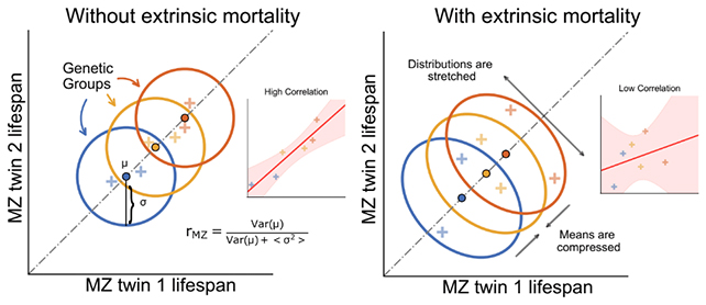
All the myriad ways in which human life can come to an end can be put into two categories: intrinsic and extrinsic. Intrinsic deaths are driven by internal factors like aging and genetics, while extrinsic deaths cover accidents, infections, and other outside causes.
In much of the historical data used in earlier studies, causes of death weren't captured in sufficient detail, making it hard to distinguish the different factors.
Here, the team analyzed data on thousands of twins, including data on siblings raised apart, which had not been considered in lifespan heritability studies before.
Twin data is crucial for genetic studies, separating the effects of genes on an individual from everything that comes after birth such as lifestyle choices, diet, and education.
Extrinsic causes of death were sifted out using mathematical models of mortality that suggest deaths are more likely to be intrinsic the older we get.
Not only did the results closely match real-world data, but the new estimate of 55 percent is also closer to existing estimates on genes accounting for variations in other aspects of our physiology, such as height.
"Such high heritability is similar to that of most other complex human traits and to life-span heritability in other species," write the researchers in their published paper.
While the new research doesn't necessarily counter earlier studies, it does suggest that the data we've used previously haven't told the full story when it comes to the balance of life and death.
Related: Something About Brazil's Oldest People May Reveal Missing Clues on Longevity
The researchers are now seeking to test their conclusions against modern datasets that do a better job of separating different causes of death. Learning that genetics has such significance in determining lifespan raises questions on which genes have the most effect and how they work – two possible areas for future research.
"If heritability is high, as we have shown, this creates an incentive to search for gene variants that extend lifespan, in order to understand the biology of aging and, potentially, to address it therapeutically," says Shenhar.
The research has been published in Science.
]]>
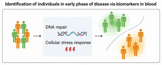
A straightforward blood test could one day reveal the earliest signs of Parkinson's disease years before more noticeable symptoms appear, according to a new study of DNA repair and cell stress.
The processes through which cells repair their DNA and adapt to stress have been linked to Parkinson's before. Here, researchers identified blood biomarkers for these mechanisms in people diagnosed with early-stage Parkinson's disease, a stage that can last for up to 20 years before the main symptoms show up.
According to researchers from Chalmers University of Technology in Sweden and the University of Oslo in Norway, the findings could inform new methods of catching Parkinson's ahead of time – and possibly finding ways to prevent it from developing.
"We highlighted biomarkers that likely reflect some of the early biology of the disease and showed they can be measured in blood," says Annikka Polster, a biostatistician at the University of Oslo.
"This paves the way for broad screening tests via blood samples: a cost-effective, easily accessible method."
As Parkinson's progresses, the death of dopamine-producing neurons reduces motor functions, thinking, and memory. In recent years, studies have shown that reductions in cell resilience and problems with DNA maintenance might be behind this damage.
For three years, the researchers tracked gene expression in blood samples in 188 healthy control participants, 393 people with fully developed Parkinson's, and 58 people with prodromal Parkinson's – the stage when the disease is just getting started in the brain.
Through comparing samples from the three groups, the study showed that variations in genes related to DNA repair and cell stress responses – and the resulting effects on blood cells – can distinguish healthy individuals from people with prodromal Parkinson's with a high level of accuracy, up to 91 percent in some cases.
Notably, markers of cell stress weren't observed in the blood of people with fully developed Parkinson's. It's almost as if the beginnings of Parkinson's trigger an emergency state in cells, which is eventually overcome by the disease.
"This means that we have found an important window of opportunity in which the disease can be detected before motor symptoms caused by nerve damage in the brain appear," says Polster.
"The fact that these patterns only show at an early stage and are no longer activated when the disease has progressed further also makes it interesting to focus on the mechanisms to find future treatments."
Once the classic motor control problems and tremors associated with Parkinson's disease start showing up, a considerable amount of damage has already been done to the brain. Preliminary tests could allow treatment and support to be put in place sooner.
The researchers estimate that it may take around five years to get a blood test like this up and running, but compared to brain scans and other Parkinson's screening techniques, taking a blood sample is simple, quick, and uncomplicated – and it's not the only blood test in development.
There are now more than 10 million people worldwide affected by Parkinson's, and we don't yet have a cure for it. The best chance of changing that might lie in catching the disease well before it's able to take hold.
Related: Missing Link Between Parkinson's Protein And Damage to Brain Cells Discovered
"By the time the motor symptoms of Parkinson's disease appear, 50-80 percent of the relevant brain cells are often already damaged or gone," says Chalmers University of Technology systems biologist Danish Anwer.
"The study is an important step towards facilitating early identification of the disease and counteracting its progression before it has gone this far."
The research has been published in npj Parkinson's Disease.
]]>
The Sun has unleashed a quartet of strong solar flares, which could herald a wild week of space weather.
It all kicked off at 12:33 UTC on February 1, when the Sun fired off an X1.0 flare.
About 11 hours later, at 23:37 UTC, a massive eruption occurred with an X8.1 flare. Two more followed on February 2, with an X2.8 flare at 00:36 UTC and an X1.6 flare at 08:14 UTC.
X-class flares are the strongest the Sun can produce. In fact, the X8.1 event was the most powerful since October 2024, and the 19th-strongest on record.

These flares erupted from a cluster of sunspots designated RGN 4366, which has only just started its Earth-facing journey, according to NOAA's Space Weather Prediction Center.
"If this spot group continues to evolve, remains complex, and erupts with any powerful solar flares, there could be increased chances of energetic particle events and possible even coronal mass ejections (CMEs) to watch for," the Space Weather Prediction Center says.
Related: The Most Violent Solar Storm Ever Detected Hit Earth in 12350 BCE
CMEs are big blasts of plasma ejected from the Sun, which are often associated with the best and worst effects of solar storms. They can produce the stunning light shows we experience as auroras – but can also disrupt satellites, power grids, and communications tech.
If it feels like strong solar activity has been in the news more than usual lately, there's a reason for that.
Our Sun has recently passed the most active phase of its 11-year cycle, which gave us some incredible light shows in 2024.
Although it's expected that solar activity should be winding down between now and the start of the next cycle, in around 2030, it seems we might still be in for some wild space weather yet.
"Forecasters expect more exciting activity," the Space Weather Prediction Center says.
]]>
Grandparents affect their grandchildren's lives in many ways, but a new study suggests seniors might benefit from this caregiving, too.
"Many grandparents provide regular care for their grandchildren – care that supports families and society more broadly," says PhD student and lead researcher Flavia Chereches, of Tilburg University in the Netherlands.
"An open question, however, is whether caregiving for grandchildren may also benefit grandparents themselves. In this research, we wanted to see if providing grandchild care might benefit grandparents' health, potentially slowing down cognitive decline."
Related: Helping Others May Be an Easy Way to Keep Your Brain Young, Study Finds
Chereches and her colleagues analyzed data from nearly 3,000 grandparents, which had initially been collected for the English Longitudinal Study of Aging.
Between 2016 and 2022, the grandparents, all over the age of 50, completed surveys about whether they had a caregiver role for their grandchildren, and if so, how frequently and in what form. They also underwent cognitive testing three times across the period.

Grandparents who were involved in caring for their grandchildren performed higher in scores of verbal fluency and episodic memory than grandparents who were not actively involved in their grandchildren's lives.
For caregiving grandmothers in particular, the benefits included a slower rate of cognitive decline across time, compared to grandmothers who weren't active caregivers.
While the study didn't compare younger and older grandparents, there were no significant differences between grandparents who gave care more or less frequently: being involved to any degree appeared to have a similar effect.
Grandparents who had a relatively high baseline of cognitive function at the start of the study were more likely to take part in activities like playing with their grandchildren and assisting them with homework, and engage in a wider variety of activities overall.
It's possible that this baseline ability could explain the trend, at least in part: older folks who start more mentally 'sharp' are going to be more able to participate in their grandchildren's lives, generally.
"Being a caregiving grandparent seemed to matter more for cognitive functioning than how often grandparents provided care or what exactly they did with their grandchildren," says Chereches. However, she emphasizes that further research is needed to see if the context of that caregiving might make a difference.
"Providing care voluntarily, within a supportive family environment, may have different effects for grandparents than caregiving in a more stressful environment where they feel unsupported or feel that the caregiving is not voluntary or a burden," she adds.
The research was published in Psychology and Aging.
]]>
An international team of scientists has discovered a record-breaking method of removing a class of harmful 'forever chemicals' from contaminated water.
Their filtration technique can mop up large amounts of per- and polyfluoroalkyl substances, aka PFAS, about "100 times faster than commercial carbon filters," claims lead author and engineer Youngkun Chung from Rice University in the US.
PFAS are synthetic substances used to protect surfaces from water, fire, and grease. Manufactured since the 1940s, they're used in raincoats, upholstery, non-stick pans, food packaging, firefighting foams, and much more.
Related: 'Forever Chemicals' in US Drinking Water Linked to Cancer, Scientists Find
They certainly proved durable: the carbon-fluorine chain at the core of these molecules is so strong, PFAS are expected to take thousands of years to break down.
Now they're in our water, soil, air, and bodies. That's a problem, because we know at least two of these 'forever chemicals' – PFOA and PFOS – are linked to cancer, cardiovascular disease, fertility issues, and birth defects.
More than 12,000 other variants remain on the market today, with largely unknown health effects.
Governments and industry are making efforts to clean up the mess, but current methods are slow and can create secondary waste.

This new filtration method uses a layered double hydroxide (LDH) material that combines copper and aluminum with nitrate.
"This LDH compound captured PFAS more than 1,000 times better than other materials," Chung says. "It also worked incredibly fast, removing large amounts of PFAS within minutes, about 100 times faster than commercial carbon filters."
The material's unique structure emerges from layers of copper and aluminum with a slight imbalance in their charge, sucking in PFOA molecules, which bind tightly with the filter.
Once the adsorption material was saturated with PFOA, the team heated the material and added calcium carbonate, which allowed them to 'clean' the LDH for reuse and strip the PFOA of its fluorine backbone, effectively destroying it.

The remaining fluorine-calcium material can be disposed into landfill safely, Rice engineer Michael Wong told The Guardian.
"We are excited by the potential of this one-of-a-kind LDH-based technology to transform how PFAS-contaminated water sources are treated in the near future," Wong says.
Though it's early days for the technology, it has already shown remarkable promise in lab studies, specifically for PFOA. The filter proved effective in tests with PFAS-contaminated water from rivers, taps, and wastewater treatment plants, and researchers hope one day it can be easily incorporated into drinking water and wastewater treatment facilities.
The research is published in Advanced Materials.
]]>
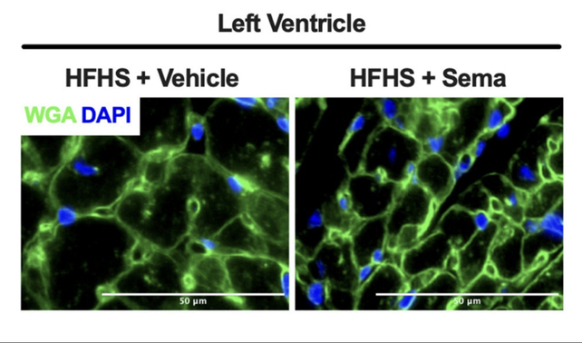
Taking oral semaglutide may reduce heart-related hospitalizations and deaths among those with a history of heart failure and type 2 diabetes, a new analysis suggests.
Data from a trial funded by Novo Nordisk – which produces the glucagon-like peptide-1 (GLP-1) receptor agonist semaglutide for weight loss (under the brand name Wegovy) and diabetes (Ozempic) – was reanalyzed by an international team of researchers.
The double-blind study involved 9,650 participants across 33 countries recruited between 2019 and 2021 and followed for almost 4 years on average.
By the end of the study period, participants with a history of heart failure had 22 percent fewer adverse cardiovascular events while taking a daily pill of semaglutide than those in a placebo group. No heart health benefits were detected in people without pre-existing heart conditions.
Related: Stopping GLP-1 Drugs Triggers Weight Regain 4x Faster Than Ending Exercise
"These data support the potential benefit of oral semaglutide in reducing heart failure events in people with type 2 diabetes and heart conditions," Oregon Health & Science University diabetologist Rodica Pop-Busui and colleagues write in their paper.
"Limitations include those intrinsic to a secondary analysis, such as the small number of participants in some of the subgroups," the team cautions.
Still, their findings are backed by previous research, which has also suggested semaglutide can reduce cardiovascular events, including strokes and heart attacks, among certain patients by almost 20 percent over about 3 years. What's more, those benefits seem to appear regardless of weight loss.
However, animal studies warn that these benefits may come with more serious risks, in addition to common side effects like nausea and vomiting.
In 2024, a study in mice revealed that semaglutide can have a shrinking effect on a type of muscle specific to mammalian hearts. Other studies have also found significant skeletal muscle loss, along with the desired fat loss and anti-diabetic effects of this molecule.
As with any medication, the use of semaglutide should be closely monitored for undesirable side effects, especially given we're still learning about its longer-term impacts.
Type 2 diabetes impacts roughly half a billion people globally, and heart failure is one of its most common complications. For people with these risk factors, semaglutide's benefits may outweigh its risks with appropriate medical guidance. However, other options, like bariatric surgery still perform better for blood sugar control than the use of this drug.
And, as we do not yet understand the mechanism behind semaglutide's impact on cardiovascular disease, researchers have called for caution when it comes to widespread prescription of this medication for purposes beyond weight loss and diabetes management.
This research was published in JAMA.
]]>
Around 10 percent of people who take statins to lower cholesterol experience mysterious muscle pains, causing many to discontinue these potentially life-saving medicines.
Now, researchers from Columbia University and the University of Rochester in the US have revealed that statin-associated muscle symptoms (SAMS), such as aches and fatigue, result from an influx of calcium into muscle cells, which leads to tissue damage and potentially life-threatening complications.
Statins work by blocking an enzyme that's required for the biosynthesis of cholesterol in the liver. As a result, levels of 'bad' LDL cholesterol are reduced in the blood, helping to prevent one of America's top killers: cardiovascular diseases like atherosclerosis, the buildup of fatty deposits in blood vessels.
Related: Our Muscles Evolved a Clever Way to Keep Us Warm, Even When They're Doing Nothing
But statins also affect "off-target" molecules, including a protein called ryanodine receptor 1 (RyR1). RyR1 is a mushroom-shaped channel, or gate, located on the sarcoplasmic reticulum, a web-like structure that surrounds muscle fibers.
RyR1 acts like a bouncer at a club, opening or closing the door to let calcium ions flow into the muscles. This calcium flow is an essential process that mediates muscle contractions.
Using mice as models, the researchers observed the precise way statins bind to RyR1, using an imaging technique called cryo-electron microscopy (cryo-EM).
Cryo-EM involves flash-freezing biological samples and then blasting them with electron beams. The deflection pattern of the electrons reveals tiny structures, allowing scientists to create highly detailed 3D images of things like proteins and view their constituent molecules.
Yet cholesterol-lowering drugs like simvastatin may keep these gates open, allowing calcium ions to leak into muscle cells, which can either directly damage muscles or trigger enzymes that degrade them.
As a result, statin users may experience persistent pain, weakness, tenderness, and cramps. The issue is exacerbated in individuals with RyR1 mutations, who may also experience episodes of malignant hyperthermia (a severe overheating triggered by medication) or weakness in the diaphragm that leads to reduced lung function and respiratory disorders.
In rare but potentially life-threatening cases, the side effects of statins can induce rhabdomyolysis, a serious syndrome in which muscle tissues rupture and leak into the bloodstream, culminating in kidney failure.
The equally gruesome autoimmune-mediated necrotizing myositis may also rarely occur, a condition in which the immune system turns against its own tissues and kills muscle tissue.
The leaky calcium gate explanation may not apply to all cases of SAMS, but now that we understand this mechanism, it could help identify people at risk of statin intolerance. Around 40 million adults take statins in the US alone, and approximately 10 percent of treated individuals experience SAMS.
"I've had patients who've been prescribed statins, and they refused to take them because of the side effects," says lead author Andrew Marks, a cardiologist at the Columbia University Vagelos College of Physicians and Surgeons.
"It's the most common reason patients quit statins, and it's a very real problem that needs a solution."
The researchers highlight two promising options. The first is to redesign statins so they don't bind to RyR1 but still inhibit cholesterol production in the liver.
Alternatively, when the researchers treated statin-intolerant mice with Rycal, an experimental class of drug used to treat patients with rare muscle diseases, they were able to close the leaky RyR1 calcium gates and prevent simvastatin-induced muscle weakness.
This research is published in the Journal of Clinical Investigation.
]]>
Screening for heart attack risk should be happening earlier for men, according to a new study that found the risk of cardiovascular disease starts climbing when men are in their mid-30s – significantly earlier than a similar trend is seen in women.
The US-based researchers behind the study followed the health of 5,112 people for an average of around 34 years. As the participants were healthy and aged 18-30 when the study started in the mid-1980s, the researchers could chart cases of cardiovascular disease (including strokes and heart failure) over time.
According to the data, 35 is the critical age when disparities between male and female cardiovascular disease risk start to appear. Most of the difference is driven by coronary heart disease (CHD), the most common cause of heart attacks, where fatty deposits clog up arteries, blocking blood flow.
Related: Human Heart Tissue Actually Can Regenerate After a Heart Attack, New Study Shows
"That timing may seem early, but heart disease develops over decades, with early markers detectable in young adulthood," says epidemiologist Alexa Freedman from Northwestern University in the US.
"Screening at an earlier age can help identify risk factors sooner, enabling preventive strategies that reduce long-term risk."
After accounting for other contributory factors, including blood pressure, cholesterol, blood sugar levels, smoking status, physical activity, and body weight, the gap was lessened – but it didn't disappear, suggesting there's more to the story.
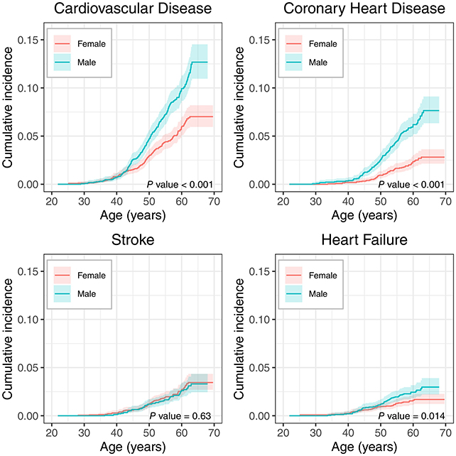
The data showed that men reach a 5 percent incidence level of cardiovascular disease about seven years earlier than women, or 50.5 years versus 57.5 years, on average. For CHD specifically, a 2 percent incidence is reached in men a decade ahead of women.
For stroke risk, there was little difference between men and women, and the gap for heart failure (where the heart isn't pumping as well as it should be) started to emerge later in life, findings that future studies may be able to build upon.
"This was still a relatively young sample – everyone was under 65 at last follow-up – and stroke and heart failure tend to develop later in life," says Freedman.
While the study didn't go into the reasons for the discrepancy between men and women in much detail, differences in sex hormones and cholesterol levels may be partly responsible.
The 10-year difference in CHD risk between the sexes has been reported before, but this new study analyzed more recent data and expanded upon previous analyses to include multiple kinds of cardiovascular disease.
Heart disease remains the leading cause of death for both men and women in the United States, and the researchers are keen not to underplay the risks for women.
However, given that women are more likely to regularly visit health professionals for check-ups, and men have such a significant head start when it comes to heart attack risk, the researchers are hoping to see more done to encourage men to get their heart health assessed at an earlier age.
"Our findings suggest that encouraging preventive care visits among young men could be an important opportunity to improve heart health and lower cardiovascular disease risk," says Freedman.
The research has been published in the Journal of the American Heart Association.
]]>
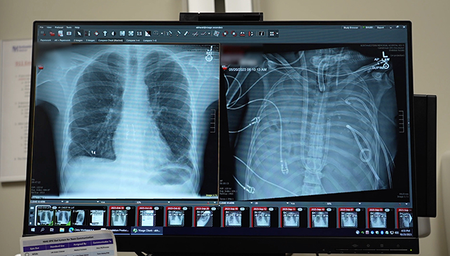
In a remarkable act of life preservation, surgeons were able to keep a critically ill man alive for 48 hours without a pair of lungs, while he waited for a double lung transplant – a radical approach that could be used again for selected patients.
A team from Northwestern University in the US built a total artificial lung (TAL) system that oxygenates blood like our lungs usually do, while managing blood flow and protecting the heart.
The TAL was crucial in stabilizing the patient and preparing him to receive a pair of donor lungs. More than two years on, the individual has recovered well – and has lungs that are fully working.
Related: Experimental Drug Helped Cancer Patients Live 40% Longer in Clinical Trial
It's a story that starts in spring 2023, when the 33-year-old man developed influenza-associated lung failure. This rapidly progressed to pneumonia, sepsis, and what's known as acute respiratory distress syndrome (ARDS).
"He had developed an infection of his lungs that just could not be treated with any antibiotics because it was resistant to everything," says thoracic surgeon Ankit Bharat.
"That infection caused his lungs to liquify and then continued to progress to the rest of his body."
The standard approach would be to put the patient on a life support system and give the lungs time to recover. Here, though, the lungs were the main problem and source of infection: The man seemed certain to die if his lungs weren't removed, and very likely to die if they were.
Removing both lungs – a bilateral pneumonectomy – usually leads to the heart failing due to disruptions in blood flow.
To avoid that and overcome the limitations of previous attempts, the medical team behind the TAL added dual blood flow channels and a flow-adaptive shunt, allowing variations in blood flow to be evened out.
The machine was enough to keep the patient alive long enough for his body to recover enough to make a lung transplant viable. Once the organs were removed, signs of recovery from the infection began.
Bharat and his team carried out a molecular analysis of the lungs after they'd been removed, confirming that there was no chance of the lungs recovering from ARDS of their own accord.
The scarring and immune damage meant that in this case, a lung transplant was absolutely necessary.
"Conventionally, lung transplant is reserved for patients who have chronic conditions like interstitial lung disease or cystic fibrosis," says Bharat.
"Currently, people think if you get severe ARDS, you keep supporting them and ultimately the lungs will get better."
This is an approach that could be used again to save more lives: While constructing a TAL system like this is currently possible only at specialized centers, Bharat hopes that the innovations applied here could be incorporated into standard devices in the future.
Whereas a double-lung transplant might previously have been considered impossible in this scenario, we now know it can be done and can be a success – and may be an option in future cases, though it still depends on timely access to donor lungs.
"In my practice, young patients die almost every week because no one realized that transplantation was an option," says Bharat.
"For severe lung damage caused by respiratory infections, even in acute settings, a lung transplant can be lifesaving."
A case report on the operation has been published in Med.
]]>
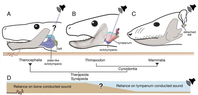
Modern mammals have unique hearing abilities, able to sense a broad range of volumes and frequencies using middle-ear features, including our eardrums and a few small bones.
A new study from paleontologists at the University of Chicago in the US has revealed these physical features began to emerge nearly 50 million years earlier than we thought.
They found their evidence within a 250-million-year-old fossil of the mammal ancestor, Thrinaxodon liorhinus. Using computed tomography scans of the animal's skull and jaw, they created 3D models that allowed them to simulate how Thrinaxodon's anatomy might have reacted to the different sound pressures and frequencies, using engineering software to see how its bones 'wiggled' in response.
Related: Our Ears Still Try to Swivel Around to Hear Better, Study Discovers
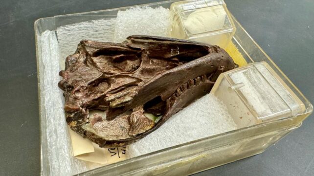
Thrinaxodon lived during the Early Triassic, before the first dinosaurs. It was a cynodont – a close relative of early mammals – with a body that looks somewhere in between a lizard and a fox.
Some of its genes follow the same blueprint modern mammals carry today, and this new study suggests the architecture of its hearing is also something we share.
Early cynodonts had ear bones – the malleus, incus, and stapes – that were attached to their jaw. In later species, these tiny fragments eventually became detached from the jaw to form the distinctly mammalian middle ear.
Before the middle ear and its associated 'tympanic' hearing abilities, animals relied on bone-conducted sound, where nerves carry signals from vibrations in the jawbone to the brain.
Paleontologists have speculated for decades that Thrinaxodon may be a 'missing link' in the evolution of mammalian hearing. In 1975, University of Wisconsin anatomist Edgar Allin proposed that Thrinaxodon might have had an early form of an eardrum stretched across the still-attached, hooked bone structure protruding from its jaw.
But at the time, Allin didn't have the technology to prove his suspicion, which is why the researchers on the new study have now revisited the question with engineering software.
"For almost a century, scientists have been trying to figure out how these animals could hear. These ideas have captivated the imagination of paleontologists who work in mammal evolution, but until now we haven't had very strong biomechanical tests," says Alec Wilken, evolutionary scientist at the University of Chicago.
"We took a high-concept problem – that is, 'how do ear bones wiggle in a 250-million-year-old fossil?' – and tested a simple hypothesis using these sophisticated tools."
The 3D model allowed the team to examine the animal's skull and jawbone in unprecedented detail, including the crook in its jawbone across which an early eardrum might have stretched.
Then, using a tool better known to engineers for testing vibrational stress on infrastructure like planes and bridges, they simulated how Thrinaxodon's skull and jaw would be affected by a range of sounds.
Of course, there's a lot more to a living head than just bone, so the scientists also filled in the gaps using known parameters from living animals about the kinds of soft tissue that might have also been at play.
"Once we have the CT model from the fossil, we can take material properties from extant animals and make it as if our Thrinaxodon came alive," says Zhe-Xi Luo, Wilken's advisor. "That hasn't been possible before, and this software simulation showed us that vibration through sound is essentially the way this animal could hear."
All together, the results suggest Thrinaxodon's eardrum would have worked quite well even without the detached middle-ear bones. And it would have been quite an upgrade on bone conduction, potentially marking a transition point for mammals towards reliance on tympanic hearing.
Wilken and team make a conservative estimate that with this built-in equipment, Thrinaxodon could have achieved a hearing range from 38 to 1,243 hertz (for reference, a healthy young person can hear around 20 to 20,000 hertz), and was most sensitive to sounds at 1,000 hertz when the sound pressure was 28 decibels (a sound level somewhere between a whisper and a normal conversation).
This would have helped Thrinaxodon locate prey, avoid predators, and may have even played a role in reproduction.
The research was published in Proceedings of the National Academy of Sciences.
]]>
An investigation by researchers from Case Western Reserve University School of Medicine in the US has filled in a missing link between the toxic build-up of proteins in the neurodegenerative condition Parkinson's disease and the death of critical brain cells.
The result of three years of research, the discovery connects alpha-synuclein proteins to a breakdown in mitochondrial function, both previously linked to Parkinson's.
"We've uncovered a harmful interaction between proteins that damages the brain's cellular powerhouses, called mitochondria," says neuroscientist Xin Qi.
"More importantly, we've developed a targeted approach that can block this interaction and restore healthy brain cell function."
Related: 'Zap-And-Freeze' Brain Imaging Could Reveal The Secrets of Parkinson's
Research has previously shown that toxic, abnormal clumps of alpha-synuclein damage neurons in Parkinson's. We also know that the disease is associated with weaker mitochondria, depriving neurons of the energy they need to work effectively.
Those two malfunctions have been linked before, but this new study provides a clearer idea of how.
In lab tests, the team observed interactions between alpha-synuclein and an enzyme called ClpP, which helps manage mitochondrial waste removal.
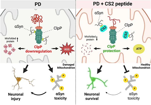
Their results suggest it is the way that alpha-synuclein binds to ClpP that disrupts mitochondria function, producing the knock-on effects so common in Parkinson's – including a decline in dopamine production.
The most significant part of the research was the development of a potential treatment to counter this damaging biochemical reaction. A short length of protein called CS2 was designed to act as a decoy for alpha-synuclein, diverting its attention away from ClpP and cell mitochondria.
In tests on human brain tissue, mouse models, and neurons developed in the lab, CS2 was shown to have positive effects. It reduced brain inflammation and restored some motor and cognitive function in animals.
"This represents a fundamentally new approach to treating Parkinson's disease," says neurophysiologist Di Hu. "Instead of just treating the symptoms, we're targeting one of the root causes of the disease itself."
Researchers estimate it might be five years until human clinical trials could evaluate CS2's safety and efficiency in humans – this kind of biological tweaking can have unintended consequences that scientists will need to extensively test for.
Nevertheless, it's a promising step forward for Parkinson's research. Not only does the study identify one of the faults that is associated with the disease at the smallest molecular level, it also demonstrates a way it can potentially be repaired.
All of this is in the context of what is already known about Parkinson's. It's a hugely complex disease where causes are difficult to differentiate from consequences – it's likely that multiple treatment approaches are going to be needed to finally find ways to cure the disease and stop it from developing in the first place.
"One day we hope to develop mitochondria-targeted therapies that will enable people to regain normal function and quality of life, transforming Parkinson's from a crippling, progressive condition into a manageable or resolved one," says Qi.
The research has been published in Molecular Neurodegeneration.
]]>
Scientists have made some intriguing parasite discoveries in an accidental back-of-the-pantry natural history museum. Canned salmon, well past its prime, has preserved decades of Alaskan marine ecology in brine and tin.
Parasites can reveal a lot about an ecosystem, since they tend to get up in the business of multiple species. But unless they cause a major issue for humans, historically we've mostly ignored them.
That's a problem for parasite ecologists, like Natalie Mastick and Chelsea Wood from the University of Washington, who had been searching for a way to retroactively track the effects of parasites on Pacific Northwestern marine mammals.
Related: 'Zombie Worms' Have Mysteriously Vanished, Troubling Scientists
So when Wood got a call from Seattle's Seafood Products Association, asking if she'd take boxes of dusty old expired cans of salmon – some dating back to the 1970s – off their hands, her answer was, unequivocally, yes.
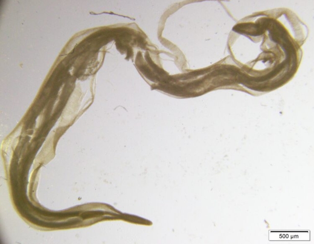
The cans had been set aside for decades as part of the association's quality control process, but in the hands of the ecologists, they became an archive of excellently preserved specimens, not of salmon, but of worms.
Watch the video below for a summary of the research:
While the idea of worms in your canned fish is a bit stomach-turning, these roughly 0.4-inch (1-centimeter) long marine parasites, anisakids, are harmless to humans when killed during the canning process.
"Everyone assumes that worms in your salmon is a sign that things have gone awry," said Wood when the research was published in 2024.
"But the anisakid life cycle integrates many components of the food web. I see their presence as a signal that the fish on your plate came from a healthy ecosystem."

Anisakids enter the food web when they are eaten by krill, which in turn are eaten by larger species.
This is how anisakids end up in the salmon, and eventually, the intestines of marine mammals, where the worms complete their life cycle by reproducing. Their eggs are excreted into the ocean by the mammal, and the cycle begins again.
"If a host is not present – marine mammals, for example – anisakids can't complete their life cycle and their numbers will drop," said Wood, the paper's senior author.
Related: Microbes in Fukushima Found Surprisingly Unscathed by Radiation
The 178 tin cans in the 'archive' contained four different salmon species caught in the Gulf of Alaska and Bristol Bay across a 42-year period (1979–2021), including 42 cans of chum (Oncorhynchus keta), 22 coho (Oncorhynchus kisutch), 62 pink (Oncorhynchus gorbuscha), and 52 sockeye (Oncorhynchus nerka).
Although the techniques used to preserve the salmon do not, thankfully, keep the worms in pristine condition, the researchers were able to dissect the filets and calculate the number of worms per gram of salmon.
They found worms had increased over time in chum and pink salmon, but not in sockeye or coho.
"Seeing their numbers rise over time, as we did with pink and chum salmon, indicates that these parasites were able to find all the right hosts and reproduce," said Mastick, the paper's lead author.
"That could indicate a stable or recovering ecosystem, with enough of the right hosts for anisakids."
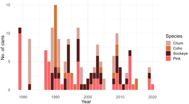
But it's harder to explain the stable levels of worms in coho and sockeye, especially since the canning process made it difficult to identify the specific species of anisakid.
"Though we are confident in our identification to the family level, we could not identify the [anisakids] we detected at the species level," the authors write.
"So it is possible that parasites of an increasing species tend to infect pink and chum salmon, while parasites of a stable species tend to infect coho and sockeye."
Related: Common Parasite Rips The Face From Your Cells to Wear as a Disguise
Mastick and colleagues think this novel approach – dusty old cans turned ecological archive – could fuel many more scientific discoveries. It seems they've opened quite a can of worms.
This research was published in Ecology and Evolution.
An earlier version of this article was published in April 2024.
]]>
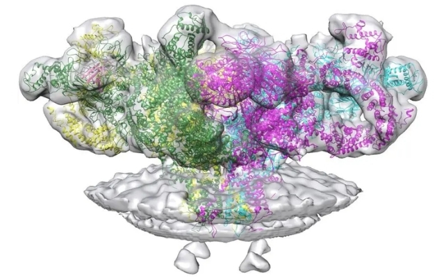
Scientists in Japan have discovered a previously unknown giant virus, offering new insight into this enigmatic category of viruses – and possibly also into the origins of multicellular life.
The virus was found infecting an amoeba in a freshwater pond near Tokyo, the researchers report. They named it "ushikuvirus" after the pond, Ushiku-numa, located in Ibaraki Prefecture.
Giant viruses were largely overlooked during the first century of modern virology, with initial discoveries often misidentified as bacteria due to their size. Yet while we barely knew they existed until recent decades, we've since learned giant viruses are all around us.
Related: Hundreds of Mysterious Giant Viruses Discovered Lurking in The Ocean
Viruses in general are considered the most abundant biological entities on Earth, and some of the most perplexing. Little is known about the evolutionary history of viruses, and there is still ambiguity about whether they qualify as living organisms.
Even if they are not living, viruses clearly wield enormous influence over all forms of life, including us. That includes not just hijacking a host's cells and causing illness, but also occasionally meddling in its evolution.
Viruses can facilitate horizontal gene transfer among living things, and some – known as retroviruses – insert their DNA into the genome of host cells. If that happens in a host's germline, viral DNA can be passed on to its offspring.
In fact, ancient retrovirus remnants now comprise up to 8 percent of the human genome, which has its perks. Retroviral DNA might have given early vertebrates the ability to make myelin, and it was key for the evolution of the placenta.
Much earlier, viruses may have sparked an even bigger, more mysterious innovation: the evolutionary leap from prokaryotes, or single-celled organisms, to eukaryotes, or multicellular organisms.
Eukaryotic cells typically have a membrane-bound nucleus, representing a "chasm in design" from their nucleus-free prokaryotic forebears. It's unclear how such a dramatic change occurred, but one intriguing theory suggests that nuclei were a gift from viruses.

Known as viral eukaryogenesis, this idea was first proposed in 2001 by Masaharu Takemura, a molecular biologist at the Tokyo University of Science. He suggested the nucleus of eukaryotic cells arose from a large DNA virus, like a poxvirus, that infected some prehistoric prokaryote.
Instead of causing trouble, the virus made itself at home in the cell's cytoplasm, eventually acquiring important genes from its host and gradually transitioning into a cellular nucleus.
This theory gained traction with the 2003 discovery of giant viruses containing DNA, which form structures called "virus factories" inside host cells. These factories are sometimes enclosed in a membrane and tend to look and function a lot like the nuclei of eukaryotic cells.
Scientists have since found a variety of these giant viruses, including species in the family Mamonoviridae and the closely related clandestinovirus, which infect certain types of amoebae. Giant viruses are highly diverse and difficult to isolate, though, so a new find like ushikuvirus is a big deal.
Takemura is still investigating viral eukaryogenesis a quarter century after he introduced the idea, and was part of the team that identified and described ushikuvirus in the new study.
"Giant viruses can be said to be a treasure trove whose world has yet to be fully understood," Takemura says. "One of the future possibilities of this research is to provide humanity with a new view that connects the world of living organisms with the world of viruses."
Ushikuvirus infects amoebae known as vermamoeba (Vermamoeba vermiformis), a habit it shares with clandestinovirus; while its shape and spiky capsid surface resemble those of medusaviruses.
It also stands out from other giant viruses, however. It forces its host cells to grow abnormally large, for example, and its capsid spikes have unique caps and fibrous structures.
Rather than preserving a host cell's nucleus and replicating inside, as clandestinovirus and medusaviruses do, ushikuvirus instead forms a viral factory and destroys the host's nuclear membrane.
These similarities and differences can be vital clues, helping scientists piece together the evolutionary history of giant viruses. Takemura and his colleagues hope to learn how and why these viruses diversified so much, as well as what role they played in the rise of eukaryotes like us.
"The discovery of a new Mamonoviridae-related virus, 'ushikuvirus,' which has a different host, is expected to increase knowledge and stimulate discussion regarding the evolution and phylogeny of the Mamonoviridae family," Takemura says.
"As a result, it is believed that we will be able to get closer to the mysteries of the evolution of eukaryotic organisms and the mysteries of giant viruses," he says.
The study was published in the Journal of Virology.
]]>
Pregnancy loss remains common around the world. About 15 percent of known pregnancies end in miscarriage, although the true number is likely much higher, since many pregnancies are lost before they are discovered.
In a new study, researchers uncovered key information about genetic factors related to aneuploidy, or an abnormality in the number of chromosomes in a cell – one of the most common causes of pregnancy loss.
A miscarriage can occur for a variety of reasons, but chromosomal abnormalities are a common factor. About half of all known miscarriages in the first or second trimester result from fetuses possessing too many or too few chromosomes.
To investigate the underpinnings of aneuploidy, researchers analyzed genetic data from nearly 140,000 in vitro fertilization (IVF) embryos, offering new details about how common genetic variations can increase some parents' risk for pregnancy loss.
"This work provides the clearest evidence to date of the molecular pathways through which variable risk of chromosomal errors arises in humans," says senior author Rajiv McCoy, a computational biologist at Johns Hopkins University.
"These insights deepen our understanding of the earliest stages of human development and open the door for future advances in reproductive genetics and fertility care," McCoy says.
Chromosomal abnormalities typically arise in the egg, with a frequency that correlates positively with a mother's age. While age is a known risk factor, our understanding of the broader genetic context has been hindered by limited data, the researchers note.
To remedy that, scientists would need to analyze a huge volume of genetic data from many thousands of embryos before pregnancy loss, along with data from their biological parents.
"This is a trait closely related to survival and reproductive success, so evolution will only allow genetic differences with small effects to be common in the population," McCoy says. "You need large samples to be able to detect these small effects."
The researchers used clinical data from pre-implantation genetic testing of IVF embryos, analyzing 139,416 embryos from 22,850 sets of biological parents in hopes of finding patterns. They identified 92,485 aneuploid chromosomes within 41,480 different embryos.
"Here the power comes from these huge sample sizes," McCoy says. "That allowed us the scale and resolution to discover several of the first well-characterized associations between the mom's DNA and her risk of producing embryos that will not survive."
The strongest of these associations was found in genes that help influence the way chromosomes pair, recombine, and gather together during the process of meiosis in egg cell lines.
A variant of the gene SMC1B, which encodes a protein that helps hold chromosomes together during meiosis, was associated with reduced crossover counts and with increased maternal meiotic aneuploidy, the study found.
The analysis also highlighted associations with several other genes involved with crossover recombination, including C14orf39, CCNB1IP1, and RNF212.
"This finding is especially compelling," McCoy says, "because the genes that emerged from our study in humans are exactly the ones that experimental biologists have detailed over decades as critical for recombination and chromosome cohesion in model organisms like mice and worms."
Female meiosis initially occurs during fetal development, as chromosomes pair and recombine, then stops for years until it resumes later in life for ovulation and fertilization.
Genetic variations may lead to problems in the interim, resulting in chromosomes that separate too readily, and thus potentially setting the stage for aneuploidy when meiosis resumes.
"Our results demonstrate that inherited differences in these meiotic processes contribute to natural variation in risk of aneuploidy and pregnancy loss between individuals," McCoy says.
Predicting individual risk for pregnancy loss will remain difficult despite these findings, the authors point out, given the importance of additional factors beyond genetics, such as maternal age and environmental exposures.
Nonetheless, understanding these genetic factors could be valuable for drug development and provides a foundation for future research into both maternal and paternal genetic variations related to pregnancy loss.
The study was published in Nature.
]]>
Manipulating how much carbon dioxide a person breathes can crank up the brain's 'sewage system', researchers have found.
The proof-of-concept study, led by neuroscientists at the University of New Mexico (UNM) and The Mind Research Network in the US, offers a tantalizing possibility for a crucial clearance system that was only identified in humans in the last decade.
The findings suggest that intermittently increasing CO2 levels in the blood could help flush toxic waste products out of the brain – maybe even preventing neurological diseases associated with these toxins, like Parkinson's or Alzheimer's.
In recent experiments with healthy participants and people with Parkinson's disease, researchers found that delivering rhythmic pulses of CO2-rich air for short periods improved the flow of their brain's waste-clearance system.
Related: Men Have Higher Risk of Parkinson's, And We May Finally Know Why
Scientists aren't yet sure why that happens, but fluctuating CO2 levels can cause blood vessels to dilate and constrict. This movement could be pushing along the nearby circulation of cerebrospinal fluid – the clear liquid behind the glymphatic system that bathes our brain and spinal cord.
Typically, as a person sleeps, hidden waves of cerebrospinal fluid gradually flush waste material from their brain. But sleep issues are commonly seen in patients with Parkinson's, possibly leading to a buildup of misfolded proteins.
Cerebral blood flow also seems less adjustable in cases of Parkinson's, and the brain typically contains greater concentrations of misfolded proteins with potentially toxic effects.
As such, some researchers have hypothesized that the brain's sewage system underlies the neurological disease.
Now, scientists want to figure out how to manipulate the glymphatic system to keep the brain fighting fit. Carbon dioxide could be a nifty way to do just that.
"We brainstormed how we could boost this response," says neuropsychologist Sephira Ryman at UNM. "That is when we realized that we could reproduce, in the awake state, the glymphatic clearing response usually linked to deep sleep using intermittent CO2."
In experiments, 63 older adults, 30 of whom had Parkinson's, underwent MRI-BOLD brain imaging while breathing in cycles of brief CO₂ elevation for about 35 seconds, followed by normal air.
This intervention, known as intermittent hypercapnia, temporarily increases blood CO₂ levels. In both healthy participants and those with Parkinson's, it altered cerebrospinal fluid flow.
In another experiment, involving 10 participants, 5 of whom had Parkinson's, participants underwent three 10-minute sessions of intermittent hypercapnia. The levels of CO₂ in their blood were then measured approximately 45, 90, and 150 minutes later.
Both healthy participants and those with Parkinson's showed increased cerebrospinal flow and glymphatic clearance. Waste products from the brain also increased in the blood, suggesting improved clearance.
One participant in the study showed evidence of amyloid-beta proteins in their blood, which is a biomarker of Alzheimer's disease. After intermittent hypercapnia sessions, their plasma levels for these toxins rose robustly.
"Intermittent hypercapnia may be able to clear peptides and proteins implicated in Alzheimer's, highlighting its potential as a disease-modifying therapy for Alzheimer's patients," the authors speculate.
Whether these changes are lasting or have a significant impact on disease pathology is unknown.
While toxins in the brain are associated with Alzheimer's and Parkinson's, it is unclear if they are active players in disease progression or merely byproducts.
Ryman and her colleagues are now investigating whether practices that focus on abdominal breathing, such as yoga, tai chi, and qigong, can also impact carbon dioxide levels and brain clearance in a similar way.
The study was published in NPJ Parkinson's.
]]>Study Finds Meat Eaters Are More Likely to Live to 100, But There's a Catch

A study of over 5,000 people in China showed that meat eaters have a higher chance of living to 100 years of age – but there are a lot of caveats.
"The study's findings may reflect the nutritional challenges of advanced age, rather than any inherent problems with plant-based diets. Crucially, this doesn't diminish the well-established health benefits of these diets for younger and healthier adults," researchers write.
Read the full story here.
Boosting One Vitamin May Have a Surprising Effect on Your Poop Schedule

New research from Spain found vitamin B1 may be a regulator of "stool frequency" – in other words, it influences how often you poop.
"Gut motility problems sit at the heart of irritable bowel syndrome, constipation, and other common gut-motility disorders," says geneticist Mauro D'Amato from the Basque Research & Technology Alliance in Spain.
"But the underlying biology is very hard to pin down. These genetic results highlight specific pathways, especially vitamin B1, as testable leads for the next stage of research, including lab experiments and carefully designed clinical studies."
Read the full story here.
If an Asteroid Hits The Moon in 2032, The Aftermath Might Reach Earth

An asteroid that might hit the Moon in 2032 would put on a stunning show for us – but could also pose a threat to people and satellites.
At its peak right around Christmas of 2032, simulations expect up to 20 million meteors per hour to hit our atmosphere, and at least on the "leading edge" of the planet, most of them with naked-eye visibility. Those would include some 100-400 fireballs (i.e., larger pieces) per hour.
Read the full story here.
Scientists Identify an Epigenetic Switch That Can Slow Production of Fat Cells

Scientists in South Korea identified a molecular switch that can slow or stop the production of fat cells – at least, in mice.
"[The study] has laid an important foundation for a more sophisticated understanding of the mechanisms behind adipocyte identity changes and, in the long term, for developing personalized treatment strategies for patients with metabolic diseases," says molecular biologist Dae-Sik Lim.
Read the full story here.
Microbes in Space Mutated And Developed a Remarkable Ability

Bacteria-hunting viruses (phages) grown in space evolved differently, potentially providing new weapons against antibiotic-resistant infections.
The team found that certain space-specific phage mutations were especially effective at killing Earth-bound bacteria responsible for urinary tract infections (UTIs). More than 90 percent of the bacteria responsible for UTIs are antibiotic-resistant, making phage treatments a promising alternative.
Read the full story here.
Scientists Found an Early Signal of Dementia Hidden in Terry Pratchett's Novels

Researchers detected signs of dementia in Terry Pratchett's works up to a decade before his diagnosis, in the form of reduced word variety.
"This was not something a reader would necessarily notice, nor did it reflect a sudden deterioration in quality. Instead, it was a subtle, progressive change detectable only through detailed linguistic analysis," the researchers write.
Read the full story here.
]]>